Diabetic Retinopathy Care
& Exams in Las Vegas
Advanced Retinal Diagnostic Mapping and Vision Preservation in Summerlin.
Diabetic retinopathy care at Precise Sight focuses on the early detection and management of retinal vascular damage caused by chronic high blood sugar. Directed by our clinical team in Summerlin, Las Vegas, NV, our practice provides advanced, high-resolution optical coherence tomography (OCT) imaging and comprehensive dilated eye exams to identify microaneurysms, control disease progression, and prevent permanent vision loss.
Understanding the Stages of Diabetic Eye Disease
Diabetic retinopathy is a serious, progressive eye condition and a leading cause of blindness among working-age adults. When blood glucose levels remain consistently elevated, it damages the delicate, microscopic blood vessels that supply oxygen and nutrients to the retina—the light-sensitive tissue layer lining the back of the eye.
As these microvessels weaken, the condition progresses through two primary physiological phases:
Non-Proliferative Diabetic
Retinopathy (NPDR):
This is the early, baseline stage of the disease where blood vessel walls begin to weaken. Microscopic bulges called microaneurysms project from the vessel structures, leaking small amounts of blood, fluid, or fatty lipids into the retinal tissue. NPDR can range from mild to severe, frequently progressing without any obvious visual warning signs.
Proliferative Diabetic
Retinopathy (PDR):
This is the advanced, highly aggressive stage of the condition. Starved for oxygen, the retina triggers abnormal growth signals that cause fragile new blood vessels to sprout across its surface. Because these new vessels are structurally unstable, they frequently rupture and leak blood into the clear, gel-like vitreous body, causing dark floaters or a sudden clouding of vision. Left untreated, PDR can cause scar tissue formation, leading to tractional retinal detachment.
The Underlying Threat of Diabetic Macular Edema
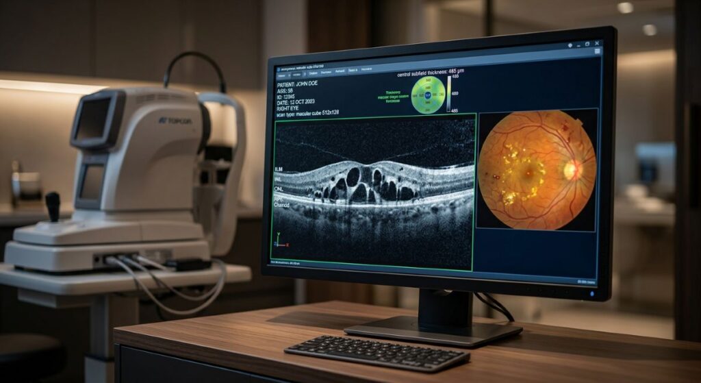
A major secondary complication that can develop during any phase of diabetic retinopathy is Diabetic Macular Edema (DME). DME occurs when the leaking fluid builds up specifically inside the macula—the highly sensitive center of the retina responsible for sharp, straight-ahead reading vision. The accumulation of fluid causes the macular layers to swell, distorting light signals and transforming mild background retinopathy into a sudden, noticeable blurring of central visual acuity.
Advanced Digital Analytics for Retinal Vascular Tracking
Because diabetic eye changes typically develop long before you note changes to your vision, relying on basic sight charts is a major clinical risk. Precise Sight utilizes advanced, university-grade digital diagnostic imaging to map your retinal circulation with exceptional accuracy:
Spectral-Domain Macular & Retinal OCT
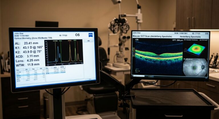
This high-speed optical coherence tomography system performs non-invasive cross-sectional laser scans of the retinal architecture. It allows Justin Hart, OD, and Eissa Hanna, MD, to look beneath the surface to find hidden micro-fluid pockets or macular swelling down to the micron.
High-Resolution Digital Fundus Photography
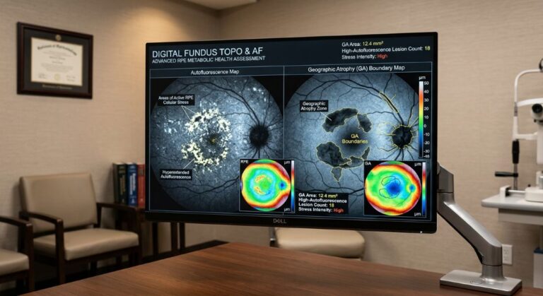
Specialized imaging that documents the surface of your retina, creating a permanent visual baseline to track changes in microaneurysms, cotton wool spots, and hemorrhages over time.
Meticulous Wide-Field Dilated Exams
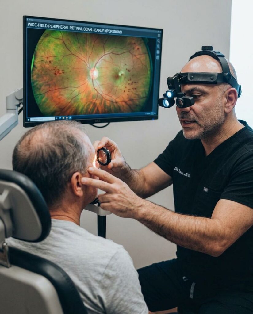
A comprehensive physical inspection of the peripheral retina to check for early vessel blockages and ensure structural integrity.
Staged Clinical Interventions and Preventive Care
Managing diabetic retinopathy is a collaborative process that scales based on your objective diagnostic tracking data:
For early to moderate NPDR, treatment centers on rigorous metabolic preservation. We coordinate closely with your primary care physician or endocrinologist to help stabilize blood glucose, hemoglobin A1c, and blood pressure metrics, slowing or stopping vascular breakdown.
If tracking metrics show a shift toward DME or Proliferative Retinopathy, our clinical network coordinates seamless transitions to premier regional retinal specialists. These targeted paths deliver micro-injections of Anti-VEGF medications or specialized laser treatments to safely shrink abnormal blood vessels and dry up macular swelling.
Quantifiable Safety Standards and Screening Milestones
Proactive annual diagnostic screenings allow our medical team to protect vision safety and predict changes for patients across the Las Vegas Valley.
95% Prevention Rate:
Severe vision loss from advanced diabetic retinopathy can be prevented through timely, proactive annual screenings and early clinical intervention.
100% Annual Standard:
The medical frequency benchmark recommended for dilated retinal tracking in all individuals diagnosed with Type 1 or Type 2 diabetes.
Sub-5 Micron Resolution:
Achieved by our in-house optical coherence tomography (OCT) systems to capture early macular structural adjustments.
A Meticulous Standard for Preserving Visual Performance
"Diabetic retinopathy does not announce its arrival with early pain or visual changes. By the time a patient notices blurriness, structural vascular damage has already occurred. At Precise Sight, we use high-resolution retinal OCT tracking to find early fluid leaks before they reach your center of vision, allowing us to proactively protect your sight and preserve your active lifestyle."
— Justin Hart, OD