Corneal Disease Treatment & Care
in Las Vegas
Advanced Diagnostics and Specialized Therapies for Complex Corneal Pathology in Summerlin.
Corneal disease treatment at Precise Sight focuses on preserving structural integrity and optical clarity of the eye’s clear front window. Managed by Harvard fellowship-trained ophthalmic surgeon Dr. Eissa Hanna and clinical optometrist Dr. Justin Hart in Summerlin, Las Vegas, NV, our boutique practice provides advanced diagnostics and custom treatments for keratoconus, Fuchs’ dystrophy, corneal scarring, and severe dystrophies.
Structural Integrity of the Cornea: Optical Clarity and Focus
The cornea is the clear, dome-shaped outer layer lining the very front of your eye. Functioning as the eye’s primary lens, it is responsible for focusing roughly 65% to 75% of the incoming light that enters your eye. To project crisp, high-definition images onto the retina, the cornea must remain perfectly smooth, uniform, and completely transparent.
Advanced Management of Progressive Corneal Disorders
Our clinical team provides university-grade diagnostics and staged medical care paths for complex corneal conditions:
1. Keratoconus (Progressive Corneal Thinning)
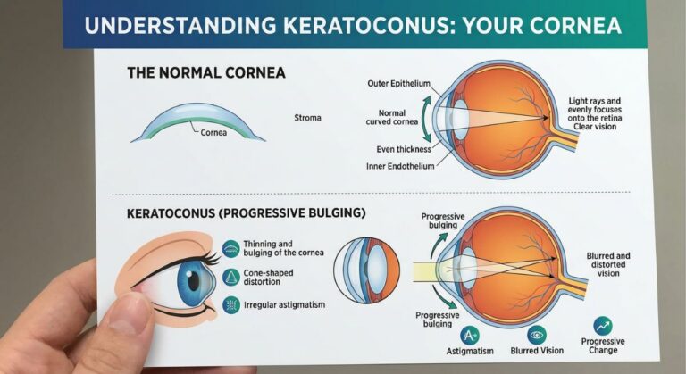
Keratoconus is a progressive ocular disease where the normally round, dome-like cornea gradually thins and bulges outward into an irregular, asymmetric cone shape. This structural warping induces severe irregular astigmatism and progressive nearsightedness.
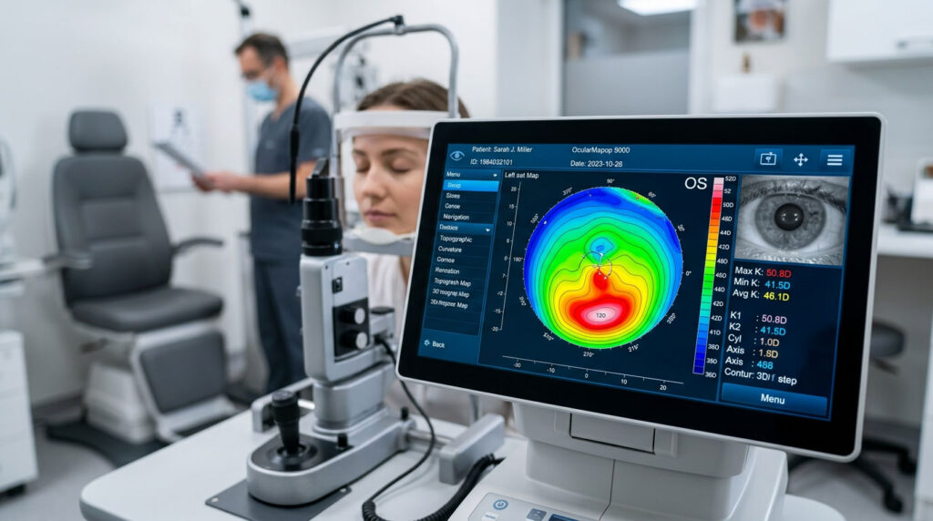
- Clinical Management: In its early stages, keratoconus is tracked using advanced corneal mapping and corrected with specialized rigid gas permeable (RGP) or scleral contact lenses. For progressive cases, we coordinate closely with regional corneal surgeons to facilitate Corneal Collagen Cross-Linking (CXL)—a specialized ultraviolet light therapy that permanently strengthens and stiffens structural corneal fibers to halt further bulging.
2. Fuchs' Endothelial Dystrophy (Fluid Accumulation)
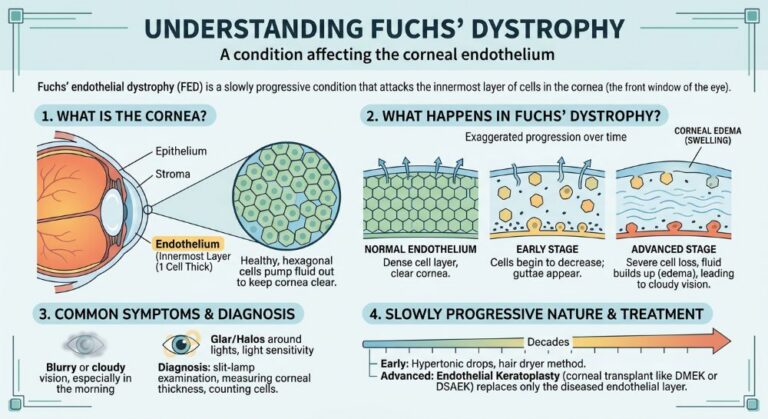
Fuchs’ dystrophy is an inherited, slowly progressive condition that attacks the innermost layer of corneal cells, known as the endothelium. In a healthy eye, these endothelial cells function as microscopic pumps, continuously drawing fluid out of the cornea to keep it thin and perfectly clear.
- Clinical Management: In Fuchs’ dystrophy, these pump cells gradually die off. Fluid accumulates inside the corneal layers, causing chronic swelling (edema), painful microscopic blisters, and a characteristic morning blurriness. Early stages are managed with hypertonic drops and ointments to draw fluid out, while advanced cases are monitored for modern endothelial keratoplasty micro-surgery.
3. Corneal Scarring, Pterygia, and Severe Dystrophies
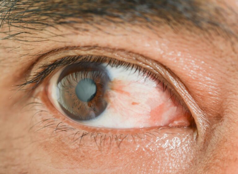
Ocular trauma, recurring infections, or advanced surfer’s eye growth can leave permanent, opaque scars across your central visual pathway. Dr. Hanna utilizes advanced anterior segment imaging to chart the exact depth of structural tissue scars, establishing if a condition can be managed conservatively or requires precision surgical intervention to restore an unhindered optical path.
High-Definition Diagnostic Technology for Corneal Mapping
Because effective treatment depends on sub-micron accuracy, Precise Sight utilizes advanced, non-invasive digital diagnostic technology. This objective data allows us to monitor corneal disease at the cellular layer:
This diagnostic system creates a highly detailed three-dimensional elevation map of both the front and back surfaces of your cornea, measuring localized curvature irregularities to track early keratoconus.
Ultrasonic and optical measurements that map the exact thickness of your corneal layers across multiple zones to identify fluid accumulation or structural thinning.
Measures the Objective Scatter Index (OSI) caused by early corneal swelling or scarring, tracing how much light scatter is occurring before it impacts your visual acuity.
Quantifiable Safety Standards and Diagnostic Milestones
Relying on precise digital metrics allows our clinical team to protect vision safety and predict structural changes for families across the Las Vegas Valley.
Over 95%
Stabilization:
Success rate achieved by Corneal Collagen Cross-Linking (CXL) in permanently halting the progression of keratoconus thinning.
500
Microns:
The average baseline safety threshold for healthy central corneal thickness, tracked meticulously via digital pachymetry.
100% Non-Invasive
Mapping:
Maintained across our advanced corneal topography suites to capture early irregular astigmatism.
A Meticulous Standard for Ocular Surface Restoration
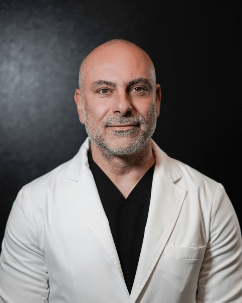
"Treating corneal disease requires absolute structural precision. The cornea is a delicate, multi-layered ecosystem that dictates how you focus on the world. By utilizing advanced 3D corneal topography alongside the HD Analyzer, we can map structural thinning or endothelial fluid buildup long before it causes permanent visual distortions, applying targeted protocols that safely preserve your clear, natural vision."
— Dr. Eissa Hanna, MD