Advanced Orbital Disorders Care
in Las Vegas

Specialized Oculoplastic and Reconstructive Orbital Surgery in Summerlin.

Orbital disorders treatment at Precise Sight focuses on diagnosing and surgically managing pathologies within the bony eye socket (orbit). Managed by Harvard fellowship-trained ophthalmic surgeon Dr. Eissa Hanna in Summerlin, Las Vegas, NV, our boutique practice provides specialized medical and reconstructive interventions for thyroid eye disease, orbital tumors, trauma-induced fractures, and inflammatory socket conditions.

Structural Dynamics of the Bony Eye Socket

The orbit is the specialized, cone-shaped bony cavity in the skull that securely houses the eyeball, its controlling extraocular muscles, crucial sensory nerves, blood vessels, and protective fat tissues. Because this bony structure is entirely rigid, any internal tissue adjustments—such as localized cellular inflammation, vascular swelling, or abnormal mass developments—instantly compromise the delicate structures within.

When pressure accumulates inside the orbit, it pushes the eyeball forward. This can lead to double vision (diplopia), restricted eye movement, severe deep-seated pain, and a progressive bulging appearance called proptosis or exophthalmos. Left unmanaged, severe orbital pressure can compress the optic nerve, threatening permanent vision loss.

Advanced Management of Complex Orbital Conditions

Our clinical team provides university-grade diagnostics and staged medical care paths for complex orbital pathologies:

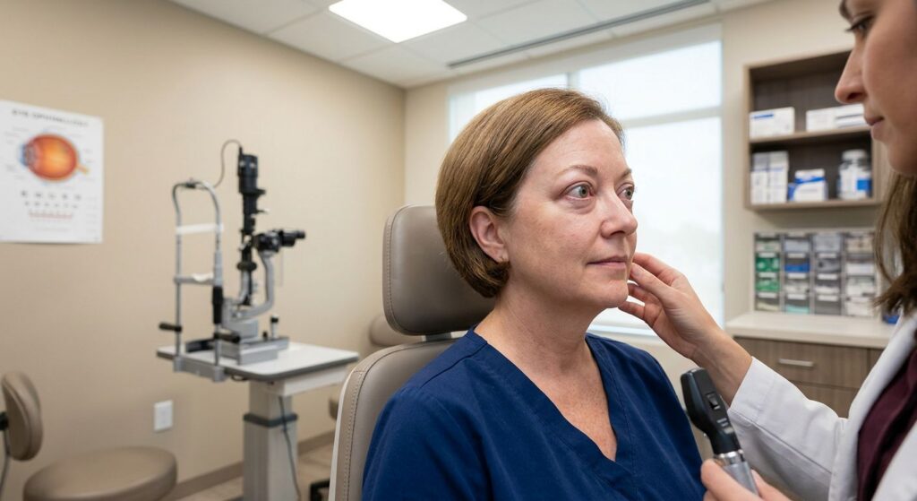
Patient in exam room with Thyroid Eye Disease

Thyroid Eye Disease is an autoimmune inflammatory disorder where the body’s immune system mistakenly attacks the soft tissues, fat, and muscles surrounding the eyes. This condition is frequently linked with systemic thyroid imbalances, such as Graves’ disease.

  • Clinical Management: TED causes severe muscle swelling and fat accumulation behind the eye, pushing the globe forward and leading to dynamic proptosis, dry eye, and painful double vision. Dr. Hanna utilizes targeted medical therapies, coordinate medical infusions, and precise structural orbital decompression surgery to expand the bony socket and safely relieve pressure when vision is threatened.
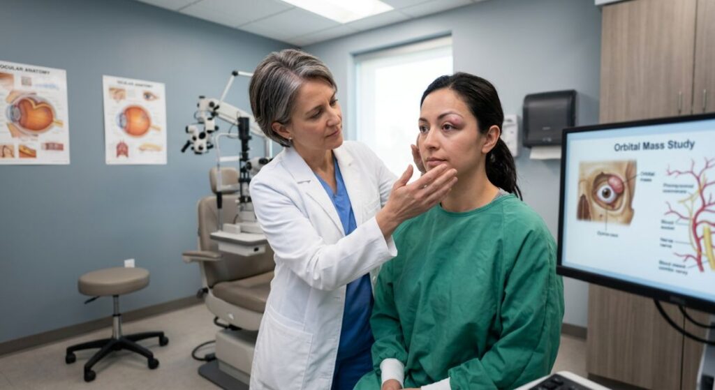
Exam room with patient who has orbital tumors and mass lesions on her eye

The orbit can host various non-cancerous (benign) or cancerous (malignant) growths, including hemangiomas, lymphangiomas, and meningiomas. These growths require exceptional diagnostic tracking.

  • Clinical Management: As an orbital tumor expands, it displaces the eye and compresses neighboring blood vessels and nerves. Utilizing detailed spatial imaging, Dr. Hanna creates a safe surgical strategy to access the orbit, perform meticulous biopsies, or completely excise the mass while protecting your eyesight.

Blunt force facial trauma can easily fracture the thin, delicate bony walls lining the floor or interior sides of the eye socket. This injury is known as an orbital blowout fracture.

  • Clinical Management: When an orbital wall fractures, the surrounding fat tissue and extraocular muscles can become physically trapped within the bony structural break. This entrapment causes severe pain and fixes the eye in place, resulting in continuous double vision when looking up or down. Dr. Hanna performs advanced reconstructive surgery, utilizing specialized microscopic biocompatible mesh plates to rebuild the natural bony floor and free trapped muscle fibers.

High-Definition Diagnostic Technology for Orbital Mapping

Because effective treatment depends on sub-millimeter anatomical mapping, Precise Sight utilizes advanced digital diagnostic technology. This objective data allows us to evaluate the deep internal structures of the eye socket:

Three Dimensional Structural Mapping

Quantifiable measurements that track the exact millimeter distance the eyeball is pushed forward out of the socket, establishing an accurate baseline for progressive thyroid eye disease.

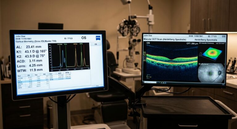
Optical Biometry and Macular OCT Scan

Optical Coherence Tomography scans the optic nerve head to ensure that increased internal socket pressure is not causing hidden nerve thinning or compromise.

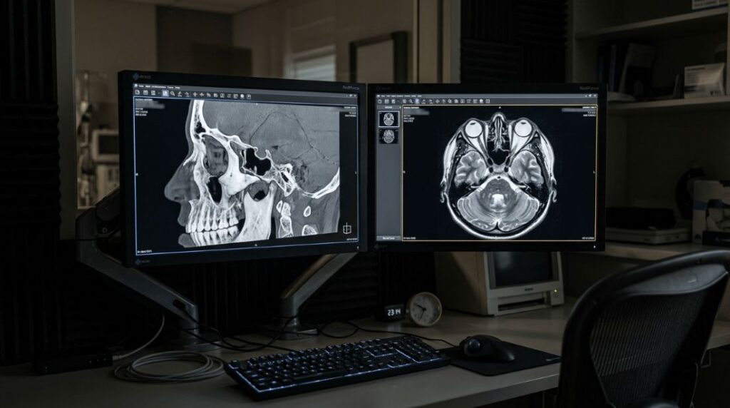
A head scan on a monitor displaying an orbital disorders diagnosis

Our clinic coordinates with top regional imaging centers to review fine-cut computed tomography (CT) scans and magnetic resonance imaging (MRI). This allows our team to inspect the bony orbit and soft tissue layers in three dimensions before planning any surgical approach.

Quantifiable Safety Standards and Reconstructive Milestones

Relying on precise digital metrics allows our clinical team to protect vision safety and predict structural changes for families across the Las Vegas Valley.

Under
2
Millimeters:

The high accuracy threshold maintained during exophthalmometry tracking to monitor structural tissue changes

0 Stitches
on the
Ocular Surface:

Achieved when using modern, transconjunctival internal incision pathways to repair orbital floor blowout fractures, completely hiding external scars.

100% Interdisciplinary Coordination:

Maintained across all auto-immune thyroid cases, linking care seamlessly with local endocrinologists.

A Fellowship-Trained Standard for Bony Socket Restoration

"Orbital disorders require a deep, highly specialized understanding of facial anatomy and neuro-ophthalmology. Because the space inside the eye socket is so limited, there is absolutely zero margin for error. Whether we are treating progressive muscle swelling from thyroid eye disease or reconstructing a blowout fracture, our priority is always to relieve structural nerve compression and restore natural symmetry, safely protecting your long-term vision."

Protect your vision
& reclaim your quality of life.